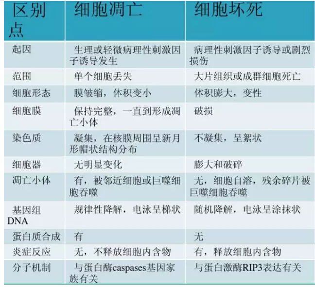
Many experimental partners have indicated that they do not know how to distinguish between apoptosis and necrosis. In fact, apoptosis and necrosis are two completely different forms of apoptosis. According to the differences in morphology, biochemistry and molecular biology of dead cells, The difference between the two.
Apoptosis refers to cell autonomy and orderly death controlled by genes for maintaining homeostasis. It involves the activation, expression and regulation of a series of genes, and is physiological and selective.
Physiological significance
Apoptosis plays an important role in embryonic development and morphogenesis, the stability of cell population in the tissue, the body's epidemic prevention and immune response, cell damage and aging caused by disease or poisoning, and the development of tumors, and has potential therapeutic significance. .
Cell necrosis
It is an abnormal physical or chemical factor or a serious pathological stimulus caused by cell damage and death, which is an abnormal death.
Cellular necrosis has long been thought to be a passive death due to pathology, but recent studies have shown that cell necrosis may be another form of "programmed death" of cells, with important physiological functions including inflammatory responses. When apoptosis does not occur properly and cells must die, necrosis is used as a "substitute" approach to apoptosis.

There are many methods for detecting apoptosis, and the commonly used morphological detection methods are described below.
1. Optical microscope and inverted microscope
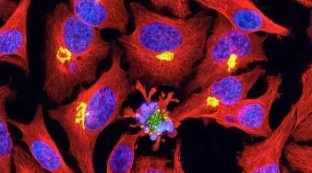
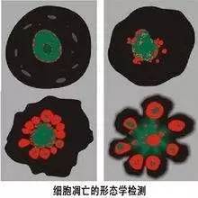
The main feature of apoptotic cells is the dense staining of nuclear chromatin, which forms dense mass and sometimes can be fragmented. In HE-stained tissue sections, the cell volume is reduced, the cytoplasm is dense, the eosinophilic staining is enhanced, and apoptotic bodies are formed. Apoptotic cells often exist in a single form in a tissue, and apoptotic cells are separated from surrounding cells without causing an inflammatory response.
The method is simple and easy to perform, but it is difficult to judge atypical cells in cell-intensive tissues, often lacks characteristic indicators, has strong subjectivity, and has poor repeatability. This method can be used for preliminary observation of the phenomenon of apoptosis, as one of the analytical indicators.
1) Unstained cells:
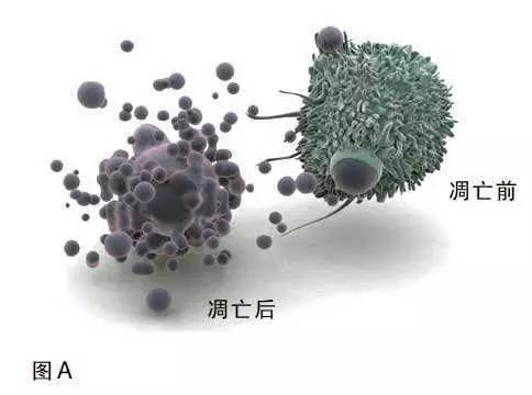
The volume of apoptotic cells becomes smaller and deformed, and the whole cell shrinks. The cell membrane is intact but foaming occurs. Apoptotic bodies are seen in the late stage of apoptosis. The apoptotic bodies are surrounded by a few round bodies around the cells. Adherent cells appear to shrink, round, and fall off.
2) Stained cells:
Giemsa staining, Wright's staining, etc.: normal cell nuclear color uniform; apoptotic cell chromatin condensation, marginalization, nuclear membrane lysis, chromatin segmentation into massive and apoptotic bodies and other typical apoptotic morphology; Necrotic cells stained lightly or not.
Hematoxylin-eosin (HE) staining: nuclear pyknosis, blue-black, cytoplasmic red (apoptotic cells), normal nucleus is evenly light blue or blue, necrotic nuclei are very pale blue or The blue color disappears.
2. Fluorescence microscopy and confocal laser scanning microscopy
The progression of apoptosis is generally judged by the morphological changes of nuclear chromatin as an indicator. Commonly used DNA-specific dyes are: Hoechst 33342, Hoechst 33258, DAPI. The combination of the three dyes with DNA is non-embedded and is primarily incorporated into the AT base region of the DNA. Bright blue fluorescence is emitted when excited by ultraviolet light.
Hoechst is a reactive dye that specifically binds to DNA and can enter normal cell membranes without much cytotoxic effect on cells. The fluorescence intensity of Hoechst 33342 in apoptotic cells is higher than in normal cells. DAPI is semi-permeable and is used for staining of conventional fixed cells.
PI and Hoechst33342 double label: PI, Hoechst33342 can bind to nuclear DNA (or RNA). However, PI cannot pass through the normal cell membrane, and Hoechst is a membrane-permeable fluorescent dye. Therefore, when the cell is necrotic or late, the cell membrane is destroyed, and the PI is red.
Both normal cells and early-stage apoptotic cells can be stained by Hoechst, but the Hoechst staining of normal nuclei is round, pale blue, with deep blue particles; while the nucleus of apoptotic cells is concentrated due to concentration. Bright blue, or nuclear lobed, fragmented, edge set. Therefore, the PI is colored as necrotic cells; the bright blue color, or the nucleus is lobulated, and the Hoechst staining of the edge set is the apoptosis cell.
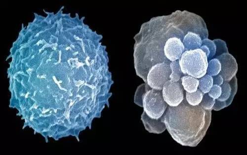
The apoptotic cells become smaller and the cytoplasm is concentrated. The morphological changes of nuclear chromatin during apoptosis are divided into three phases: the nucleus of stage I is rippled or creased, and some chromatin is concentrated; the chromatin height of phase IIa nucleus Condensation and marginalization; the nuclear cleavage of phase IIb is fragmentation, producing apoptotic bodies.

3. Electron microscope
Although the phenomenon of membrane blistering and apoptotic bodies can be seen under the light microscope, the morphological changes of apoptotic cells mostly occur in the ultrastructure, so it is difficult to observe with light microscopy, and the transmission electron microscope can clearly observe Changes in cellular structure during different periods of apoptosis. Electron microscopic observation is the most classic and reliable method for judging apoptosis, and is considered to be the gold standard for determining apoptosis.
Detection method:
Transmission electron microscopy specimens were fixed by glutaraldehyde and hungry acid, acetone dehydrated, epoxy resin embedded, ultrathin section, acetic acid tannic acid and heavy staining, observed by transmission electron microscopy. Scanning electron microscope specimens were fixed by glutaraldehyde and hungry acid, ethanol was dehydrated step by step, CO2 critical point was dried, vacuum sprayed gold, and observed by scanning electron microscopy.
Result evaluation:
It can be seen that the microvilli on the surface of apoptotic cells disappeared, nuclear chromatin condensation, marginal collection, often crescent-shaped, nuclear membrane wrinkles, tight cytoplasm, organelle concentration, membrane blistering or "bud" and small apoptosis The body and apoptotic bodies are engulfed by adjacent macrophages.
Disadvantages:
1) can only be qualitative, not quantitative;
2) The processing of specimens is complicated, the equipment is relatively expensive, and the technical level of the inspectors is relatively high, which is not suitable for large-scale specimen testing;
3) When electron microscopic observation is performed on the tissue section, sometimes apoptosis is difficult to distinguish from normal cell mitosis, because chromatin condensation can occur in both cases. If fluorescent staining is performed at the same time, it can make up for the shortage of electron microscopy.
The quality appears in a concentrated state; the chromatin in the phase IIa nucleus is highly coagulated and marginalized; the nuclear cleavage in the phase IIb is fragmentation, resulting in apoptotic bodies.
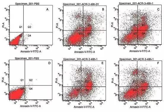
4. Flow cytometry
Flow cytometry detects apoptotic cells by examining their light-emitting characteristics and fluorescence parameters. When the cells pass through the laser beam collection point of the flow cytometer, the laser is scattered, and the analyzed scattered light can provide information on the size and structure of the cells. Since light scattering is not a specific indicator of apoptotic cells, mechanical damage and necrosis of cells can also attenuate forward scattered light. Therefore, apoptotic cells can be accurately identified only by combining the detection of light scattering properties with the detection of fluorescence parameters.
During the process of apoptosis, the endonuclease degrades between the nucleosomes of DNA molecules, causing leakage of small molecules of DNA, and the content of nuclear DNA is decreased. After flow cytometry analysis, the cells can be found to be normal on the DNA histogram. A subdiploid peak (xub-G1 peak, AP peak-apoptotic peak) appears before the Go/G1 peak of ploidy cells, representing apoptotic cells.
The morphological detection of apoptosis is intuitive and is still the gold standard for determining whether cells undergo apoptosis. However, it is time-consuming and laborious to observe the morphological characteristics of apoptotic cells, which is not suitable for the detection of a large number of apoptotic samples.
Friends who are interested in or have problems with cell culture can join
"CellMax Cell Culture Exchange Group: 515831066 "
Everyone can discuss each other's exchanges!
Or
Pay attention to the Cellmax service number (public number)


Head Protective Cap,Simplex Work Cap,Protective Cap,Anti Smashing Safety Helmet
Dongying Hong Xing Labor Protection Products Co.,Ltd. , https://www.hongxinglabor.com